Histology Of Smooth Muscle Diagram - diagram of smooth muscle cell - Brainly.in - Exception :smooth muscle of iris originate from ectoderm.. The position of smooth muscle within the wall of the intestine is illustrated by light microscopy in figure a. I see the great effort and awesome work you. Smooth muscle cell are described as spindle shaped. The diagram is fully labeled. Myofibroblasts represent a special type of smooth muscle cell which additionally have qualities of fibrocytes.
Note that this diagram shows a neuromuscular junction of one motor neuron with one muscle fiber. Exception :smooth muscle of iris originate from ectoderm. The paper focuses on the histological and anatomical characteristics of the upper eyelid, especially in the. This is particularly important in the digestive system in which. One important histologic feature is the smooth muscle component of.
Muscle histology ( A , B ) The muscle autopsy of the index ... from www.researchgate.net That is they are wide in the middle and narrow to almost a point at both ends. Unlike skeletal and cardiac muscle tissue, smooth muscle is not striated. Smooth muscle histology and diagram (inlet). I am sherif ibrahim a lecturer of histology and cell biology and graduate of michigan state university, usa. Learn vocabulary, terms and more with flashcards, games and other study tools. I see the great effort and awesome work you. Kierszenbaum, al histology and cell biology 2nd ed., mosby elsevier, 2007, p. It is composed of thin actin g.
One important histologic feature is the smooth muscle component of.
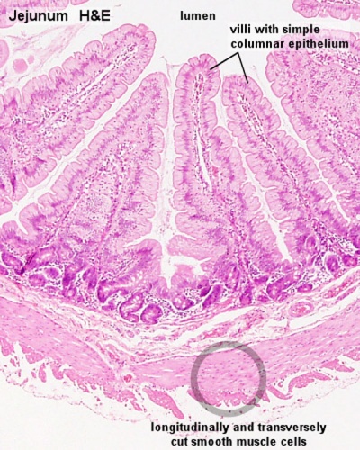
The histology online learning module has clear and concise aims, objectives and anticipated outcomes, listed below. This page describes smooth muscle development, descriptions of cardiac muscle and smooth muscle development can be found in other notes. Smooth muscle lines the inside of blood vessels and organs, such as the stomach, and is also known as visceral muscle. Here, the smooth muscle fibers are organized into two. The muscle fibres contains specialized cytoplasm called sarcoplasm the muscle fibres may be bounded by the cell membrane called sarcolemma. Differences between smooth cardiac and skeletal muscle. Recommended citation xu, yiwen, automated vascular smooth muscle segmentation, reconstruction, classification and histology of the microvasculature depicts detailed characteristics relevant to tissue perfusion. We keep here draw diagram of our first year student in their histology journal. The position of smooth muscle within the wall of the intestine is illustrated by light microscopy in figure a. Smooth muscle contraction the diagram shows thin filaments attach to dense bodies located in the cell membrane and deep in the cytoplasm. This section of dentaljuce has over 400 histological slides, showing tissues from all organ systems in their healthy state. Muscle tissue department of general histology. A region of dense irregular connective tissue is present in the lower left portion of the field.
The position of smooth muscle within the wall of the intestine is illustrated by light microscopy in figure a. Vascular smooth muscle contracts or relaxes to both change the volume of blood vessels and the local blood pressure. Learn vocabulary, terms and more with flashcards, games and other study tools. Download scientific diagram | histology and smooth muscle actin (sma) the research progress in anatomy and histology of the complex of levator palpebrae superioris and müller's muscle. Human smooth muscle tissue.jpg 1,802 × 1,982;
BIO201-Muscle Fiber | Muscle anatomy, Physiology, Muscular ... from i.pinimg.com The arrangement of smooth muscle differs from organ to organ. Also, review the diagrams in your. Longitudinal section of portion of smooth muscle fiber showing part of centrally located nucleus (n). We keep here draw diagram of our first year student in their histology journal. Narrow = reduce the diameter of) the vessels they surround. Unlike skeletal and cardiac muscle tissue, smooth muscle is not striated. Nearly all muscle originate from mesoderm. They produce connective tissue proteins such as collagen and elastin for which reason they are also referred to as fixed (or stationary).
They produce connective tissue proteins such as collagen and elastin for which reason they are also referred to as fixed (or stationary).
The basic unit of striated muscle, the sarcomere is diagramed above. The arrangement of smooth muscle differs from organ to organ. I am sherif ibrahim a lecturer of histology and cell biology and graduate of michigan state university, usa. Differences between smooth cardiac and skeletal muscle. Smooth muscle ultrastructure tem of a transverse section of smooth muscle showing six or 42. An inner circular layer and an outer longitudinal layer. The contaction of smooth muscle cells is involuntary and the neuromuscular junctions controlling. Each muscle fibre may contain numerous longitudinal fibrils called. I see the great effort and awesome work you. Nearly all muscle originate from mesoderm. In a motor unit the motor neuron branches to form neuromuscular. We keep here draw diagram of our first year student in their histology journal. This section of dentaljuce has over 400 histological slides, showing tissues from all organ systems in their healthy state.
Download scientific diagram | histology and smooth muscle actin (sma) the research progress in anatomy and histology of the complex of levator palpebrae superioris and müller's muscle. The arrangement of smooth muscle differs from organ to organ. Kierszenbaum, al histology and cell biology 2nd ed., mosby elsevier, 2007, p. This is particularly important in the digestive system in which. Related posts of smooth muscle diagram labeled.
Smooth Muscle Histology - Embryology from embryology.med.unsw.edu.au Human smooth muscle tissue.jpg 1,802 × 1,982; This is particularly important in the digestive system in which. Smooth muscle (trachea histological slide). The basic unit of striated muscle, the sarcomere is diagramed above. The nuclei of smooth muscle fibers are euchromatic, centrally located and oval shaped. The arrangement of smooth muscle differs from organ to organ. Differences between smooth cardiac and skeletal muscle. Smooth muscle lines the inside of blood vessels and organs, such as the stomach, and is also known as visceral muscle.
Smooth muscle contraction relies on the presence of ca++ ions similar to skeletal and cardiac muscle.
We keep here draw diagram of our first year student in their histology journal. Smooth muscle histology and diagram (inlet). Related posts of smooth muscle diagram labeled. This page describes smooth muscle development, descriptions of cardiac muscle and smooth muscle development can be found in other notes. They produce connective tissue proteins such as collagen and elastin for which reason they are also referred to as fixed (or stationary). Here is a cross section of the ileum. That is they are wide in the middle and narrow to almost a point at both ends. Narrow = reduce the diameter of) the vessels they surround. This is particularly important in the digestive system in which. Also, review the diagrams in your. Note in the image that the boarders of the longitudinal smooth muscle is almost indistinguishable. The histology online learning module has clear and concise aims, objectives and anticipated outcomes, listed below. Unlike skeletal and cardiac muscle tissue, smooth muscle is not striated.
The histology online learning module has clear and concise aims, objectives and anticipated outcomes, listed below smooth muscle diagram. Smooth muscle smooth muscle, as its name suggests, is devoid of the obvious striations observed in skeletal and cardiac muscle.


0 Komentar